
Women's Care, Cancer
What is contrast enhanced mammography?
Key Takeaways:
- In addition to mammography, breast ultrasound and breast MRI, another tool for finding breast cancer is contrast enhanced mammography (CEM)
- CEM is similar to traditional mammography but involves the use of IV-administered contrast
- Not everyone is a candidate for CEM, but is best for patients with dense breast or who have a high-risk of getting breast cancer
- Contrast enhanced mammography is available at the Telfair Pavilion at Candler Hospital.
St. Joseph’s/Candler offers this additional screening tool at the Telfair Pavilion to help detect breast cancer in certain patients
St. Joseph’s/Candler offers another beneficial tool to its already loaded arsenal for breast cancer screening. It’s called contrast enhanced mammography, or CEM.
CEM is performed following a screening mammogram to further look for any possible abnormal findings. It does not replace traditional mammography but can serve as a beneficial supplemental option, especially for women with dense breasts.
St. Joseph’s/Candler added CME as part of our tireless commitment to improving breast cancer detection rates. We know when breast cancer is found early, it is much more treatable and curable. CEM is currently offered at the Telfair Pavilion at Candler Hospital.
What is contrast enhanced mammography?
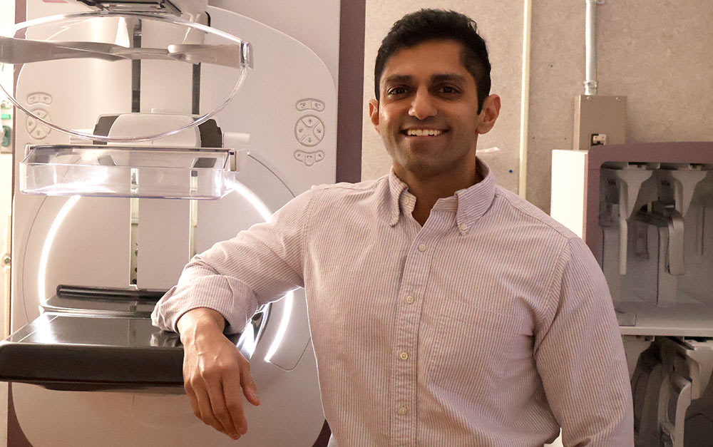
CEM very similar to a traditional mammogram, says Dr. Dil Patel, radiologist and Medical Director of the Telfair Pavilion. It just involves one extra step – IV-administered contrast.
“We still get the traditional mammogram images, and then we’ll get another set of images after we inject the contrast,” Dr. Patel explains. “On those images, if there is anything abnormal, anything hyper vascular that takes up a lot of blood, it will show up as a bright white spot against the background of dark gray tissue. The contrast helps us, as radiologists, pick up things easier.”
CEM is not for every woman. It’s best in a certain subset of patients such as those with dense breast or who have a high-risk of getting cancer, Dr. Patel says. CEM also can serve as additional screening for those who need a breast MRI, but can’t get one because they have a pacemaker or are claustrophobic.
“This doesn’t replace screening mammogram or breast MRI. I think those are excellent tools we have,” Dr. Patel says. “But, this is a nice tool that can supplement those for the patients who qualify for contrast enhanced mammography.”
Related Article: Mammography 101: Mammograms, self-exams and when to start
The patient experience
If you need CEM in addition to your screening mammogram, the good news is it’s done during one appointment. After you check-in, the nurse will place the IV and calculate the dosing of contrast, which is based on weight, Dr. Patel explains. The radiologist confirms dosing is correct and everything is OK to get started.
Typical mammogram images are taken first. The CC View, or cranial caudal, comes down from the top (cranial) all the way down to the caudal (meaning feet). The second view is called MLO, or mediolateral-oblique, and comes from the side at an angle.
Next the contrast is administered through the IV and more images of the breasts are taken. The radiologist looks at those images in real time and determines if any additional views are needed.
After all the images are taken, the patient is free to go home. You may be advised to drink plenty of water and stay hydrated to flush the contrast out of your system.
So, is it safe?
Simply put, yes. Dr. Patel says most patients tolerate the IV contrast very well. Your kidney function is monitored during the screening, especially if you have kidney issues or diabetes. If you do have any type of reaction to the contrast, which is very rare, the team has medications on hand and is prepared to manage your response.
“It’s a very low dose of radiation and the IV contrast is very well tolerated in most patients so there’s low risk to that,” Dr. Patel says. “We’ve been using contrast with MRI and CT for a long time and patients do fine with it. Certainly the benefits outweigh the minimal risks that the patient would undertake for this.”
Another breast cancer diagnostic tool: contrast enhanced biopsy
If imaging reveals an area needs to be sampled to determine whether benign or cancerous, St. Joseph’s/Candler also offers contrast enhanced biopsy. This process is similar to traditional stereotactic biopsy, which is also performed through the mammography machine.
The difference, again, is contrast is used to light up the targeted area for biopsy, Dr. Patel says.
Contrast enhanced biopsy also is available at the Telfair Pavilion. It’s only utilized when there’s a suspicious finding from a CEM, Dr. Patel says. So if there’s something abnormal seen on a traditional screening mammogram, you’d get traditional stereotactic biopsy, for example. Or, if you had a screening ultrasound, you’d most likely get an ultrasound biopsy. This makes it easier on the patient, Dr. Patel says.
If you are interested in CEM, talk to you referring healthcare provider or call the Telfair Pavilion at 912-819-7000.